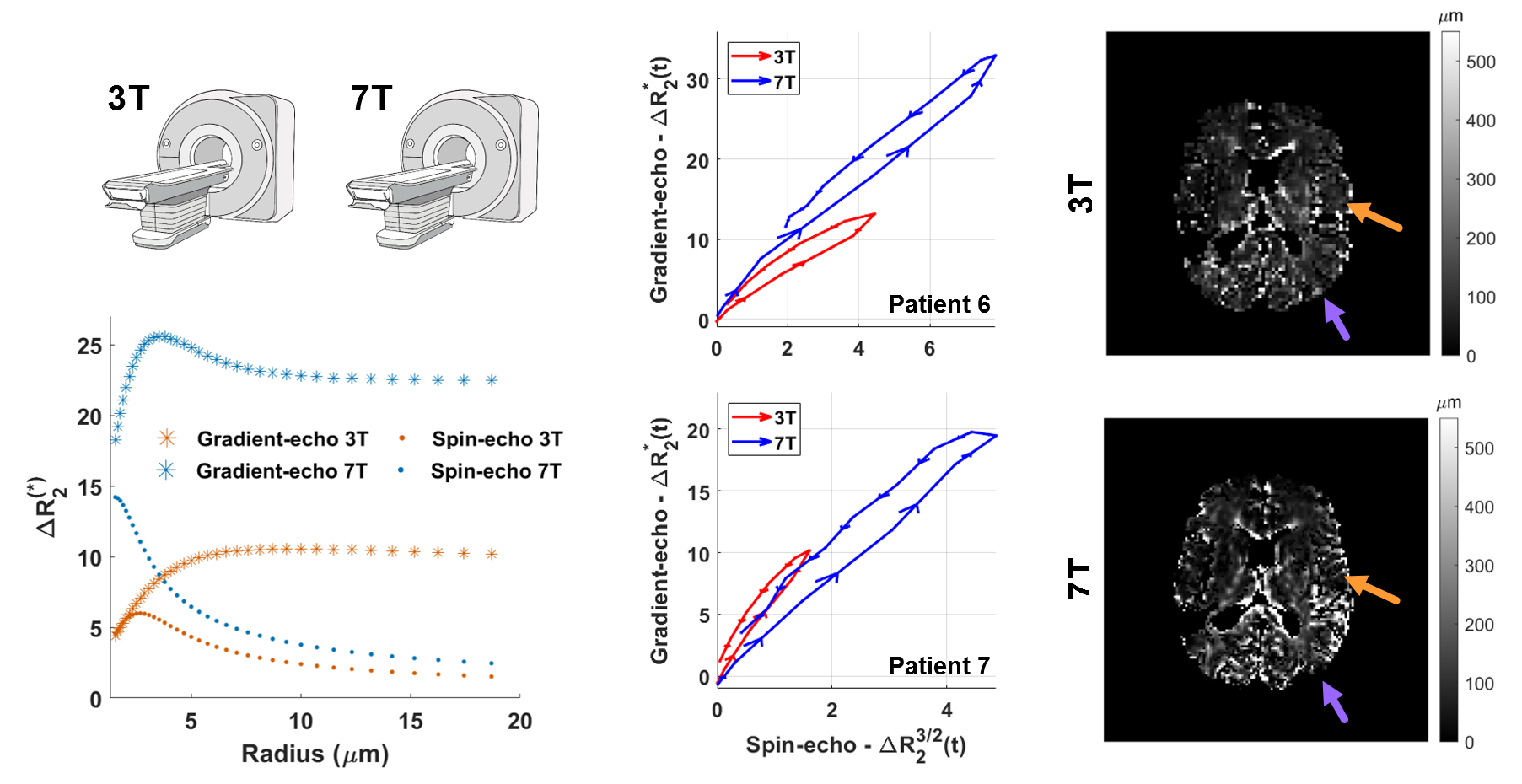
In this project, Karen van der Werff developed a protocol for spin- and gradient-echo DSC-MRI at 7T; a technique that allows to characterize the microvasculature in the brain (think of blood volume, vessel size and architecture), with the potential of improved resolution and contrast at higher field strength. Using both simulations and in vivo data from patients with glioma, she found that it is possible to probe even smaller vessels at 7T while improving image resolution and, perhaps most importantly, reducing the contrast agent dosage needed.
The open-access article can be found here.