About the research topic
In surgery of the lower spinal column, the surgeon needs to locate the correct level for the surgical intervention on the patient without direct vision on the spinal column. Current approaches for determining this level are not always accurate, and may require an X-ray system, which is cumbersome and uses ionizing radiation. In this project we develop an innovative approach where ultrasound imaging, a harmless and relatively cheap imaging modality, is used to determine in real-time the correct level for the surgery. The essence is a automated, quantitative measurement of differences in lateral profiles of spinous processes. To this end, we developed:
- an automated bone segmentation approach for segmenting the spinous process in 2D ultrasound images,
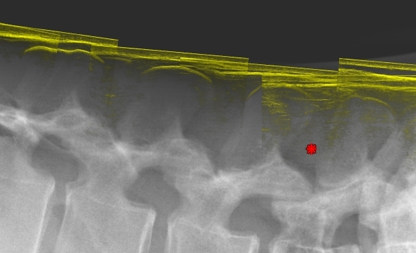
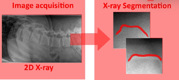
- an automated bone segmentation approach for lumbar spinal column X-ray images,
- a shape-based matching approach that determines the vertebral body level based on the shapes from X-ray and ultrasound images.
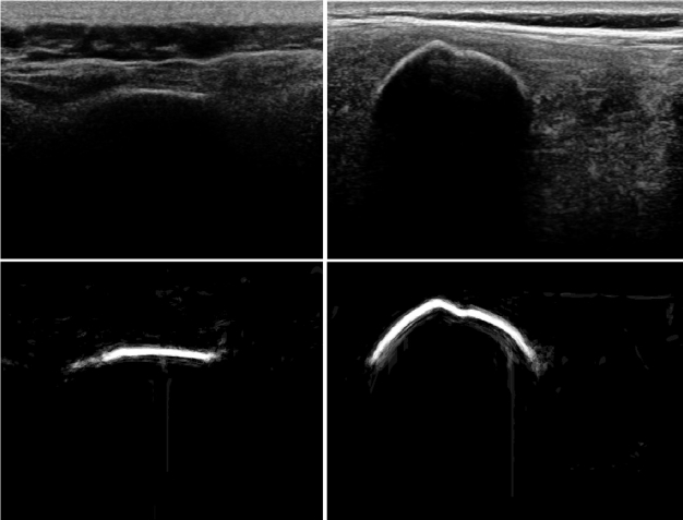
Baka et al., UMB 2017 : Ultrasound (US) imaging is a safe alternative to radiography for guidance during minimally invasive orthopedic procedures. However, ultrasound is challenging to interpret because of the relatively low signal-tonoise ratio and its inherent speckle pattern that decreases image quality. Here we describe a method for automatic bone segmentation in 2-D ultrasound images using a patch-based random forest classifier and several ultrasound specific features, such as shadowing. We illustrate that existing shadow features are not robust to changes in US acquisition parameters, and propose a novel robust shadow feature. We evaluate the method on several US data sets and report that it favorably compares with existing techniques. We achieve a recall of 0.86 at a precision of 0.82 on a test set of 143 spinal US images.
 Roodhorst, MSc project TUE :
Purpose of this project was to automate the X-ray spinous process segmentation,
and investigate the effect of the segmentation automation on the overall
localization accuracy. We implemented a U-net-based segmentation for X-ray
spinous process contours. Using manual localization and automatic segmentation
using U-net, results show a X-ray spinous process segmentation accuracy of 80%
DSC similarity.
Roodhorst, MSc project TUE :
Purpose of this project was to automate the X-ray spinous process segmentation,
and investigate the effect of the segmentation automation on the overall
localization accuracy. We implemented a U-net-based segmentation for X-ray
spinous process contours. Using manual localization and automatic segmentation
using U-net, results show a X-ray spinous process segmentation accuracy of 80%
DSC similarity.
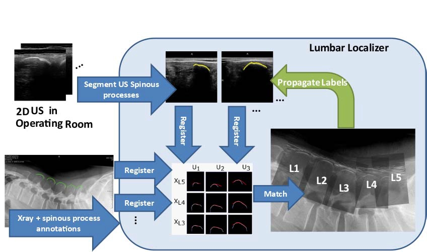
Baka et al., TMI 2017 : Localization of the correct vertebral level for surgical entry during lumbar hernia surgery is not straightforward. In this paper, we develop and evaluate a solution using free-hand 2-D ultrasound (US) imaging in the operation room (OR). Our system exploits the difference in spinous process shapes of the vertebrae. The spinous processes are pre-operatively outlined and labeled in a lateral lumbar X-ray of the patient. Then, in the OR the spinous processes are imagedwith 2-D sagittalUS, and are automatically segmented and registeredwith the X-ray shapes.After a small number of scanned vertebrae, the system robustly matches the shapes, and propagates the X-ray label to the US images. The main contributions of our work are: we propose a deep convolutional neural network-based bone segmentation algorithm from US imaging that outperforms state of the art methods in both performance and speed. We present a matching strategy that determines the levels of the spinal processes being imaged. And lastly, we evaluate the complete procedure on 19 clinical data sets from two hospitals, and two observers. The final labeling was correct in 92% of the cases, demonstrating the feasibility of US-based surgical entry point detection for spinal surgeries.