A new paper by the Eye Image Analysis Group (EyeR) including Sander, Theo, Danilo, and Luisa from the BIGR has now been published in Ophthalmology Science!
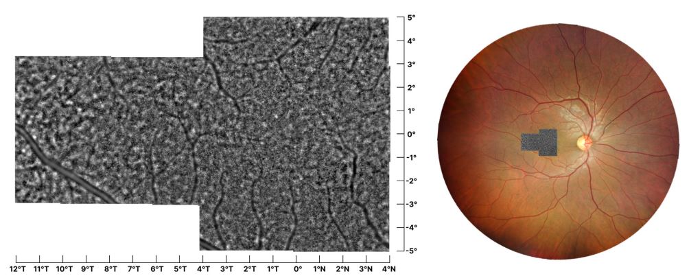
This study proposes a novel methodology on detecting cone photoreceptor cells in Adaptive Optics Flood Illumination Ophthalmoscopy (AO-FIO) using the rtx1 AO retinal camera. In particular, they develop and validate AO-FIO ConeDetect, a deep learning model for automated cone photoreceptor detection in AO-FIO images at various eccentricities and over larger regions.
This collaborative work by the renowed universities (including UMC) from the Netherlands, Belgiumm, Australia, and Switzerland as well as Imagine Eyes of France found that the performance of the developed deep learningebased model, AO-FIO ConeDetect, was comparable to that of graders from 3 medical centers. It outperformed the manufacturers’ software auto-detection.
For years, conventional imaging methods like fundus photography and OCT have struggled to detect subtle retinal changes, affecting both patient outcomes and clinical efficiency. This not only delays diagnosis but also places a burden on clinics that are often understaffed.
Recent AI advancements, combined with high-resolution imaging like AO-FIO, offer a powerful and cost-effective way to detect and quantify these changes. As shown in our study, there is still work to be done—not only in improving imaging interpretability but also in optimizing and establishing imaging protocols for clinical care.
As a community, it is our mission to drive these innovations forward, contributing to better diagnostics and prognostics of retinal diseases.