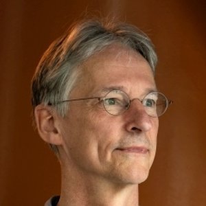
Theo van Walsum
Professor / BIGR Treasurer, Erasmus MC
Interests
- Medical Imaging
- Image Guidance and Navigation
- Visualization
- Augmented Reality
- Software Engineering
Education
-
MSc Computer Science, 1990
Delft University of Technology
-
PhD, 1995
Delft University of Technology
Research lines
Biography
Theo van Walsum graduated in Informatics (Computer Science) at the Delft University of Technology in 1990. In 1995 he received his PhD at Scientific Visualization Group of Delft University of Technology. Subject of his thesis was flow visualization on curvilinear grids. From June 1995 to May 1996, he was a research fellow at the Laboratory of Clinical and Experimental Image Processing (LKEB), at the University Hospital Leiden, where he worked on visualization of MRI and MRA data. From June 1996 to January 2005, he worked as a Postdoc at the Image Sciences Institute at the UMC Utrecht, on simulation and visualization for endovascular treatment of AAA, the image processing software environment, catheter tracking for image guidance and image guidance using 3D Rotational X-Ray imaging. In 2005 he moved to the Biomedical Imaging Group Rotterdam at the Erasmus MC.
Since 2005, he is heading the “Image Guidance in Interventions” theme group at the Biomedical Imaging Group Rotterdam. This group focusses on improving image guidance by integrating pre-operative image information in various interventional procedures. Challenges addressed are the modeling and tracking of motion and deformation of the anatomy, and the instruments. Such trackerless navigation approaches have been implemented for ultrasound and x-ray guided procedures such as TIPS, TACE and ablation of liver lesions. Currently, this research is extended with the integration of augmented reality devices to integrate the information in the direct view of the clinician. Additionally, he is involved in the cardiovascular image processing theme group, focussing quantitative imaging biomarkers for the heart and coronaries (mainly CT-based) and for stroke (CTA, DSA).
He co-organized three cardiovascular “Grand Challenges” that were organized by the BIGR, the coronary artery tracking challenge (coronary.bigr.nl), the carotid lumen segmentation and stenosis grading challenge (cls2009.bigr.nl), and the coronary stenoses detection and quantification challenge (coronary.bigr.nl/stenosis). He also initiated the MICCAI SWITCH workshop series that aims to bring together clinical and medical imaging experts working in the field of stroke and stroke treatment.
He is also active in the CONTRAST consortium (archiving of trial and registries imaging data), and is one of the leaders of the PP CONTRAST TKI program funded by Health Holland.