Biomedical Imaging Group Rotterdam
The Biomedical Imaging Group Rotterdam (BIGR) is at the forefront of research in medical image analysis & artificial intelligence (AI). We aim to improve efficiency and quality of healthcare by developing innovative AI methods in medical imaging.
We focus on both fundamental and applied research, covering the topics of image analysis, machine learning, image reconstruction, quantitative imaging biomarkers, image-guided interventions, making use of both research data and routine clinical data.
We have a strong outward look: towards other imaging sources, other diagnostic modalities, integrated diagnostics, collaboration with clinical departments. We have strong collaborations with many researchers, clinicians and industry partners.
March 9, 2026
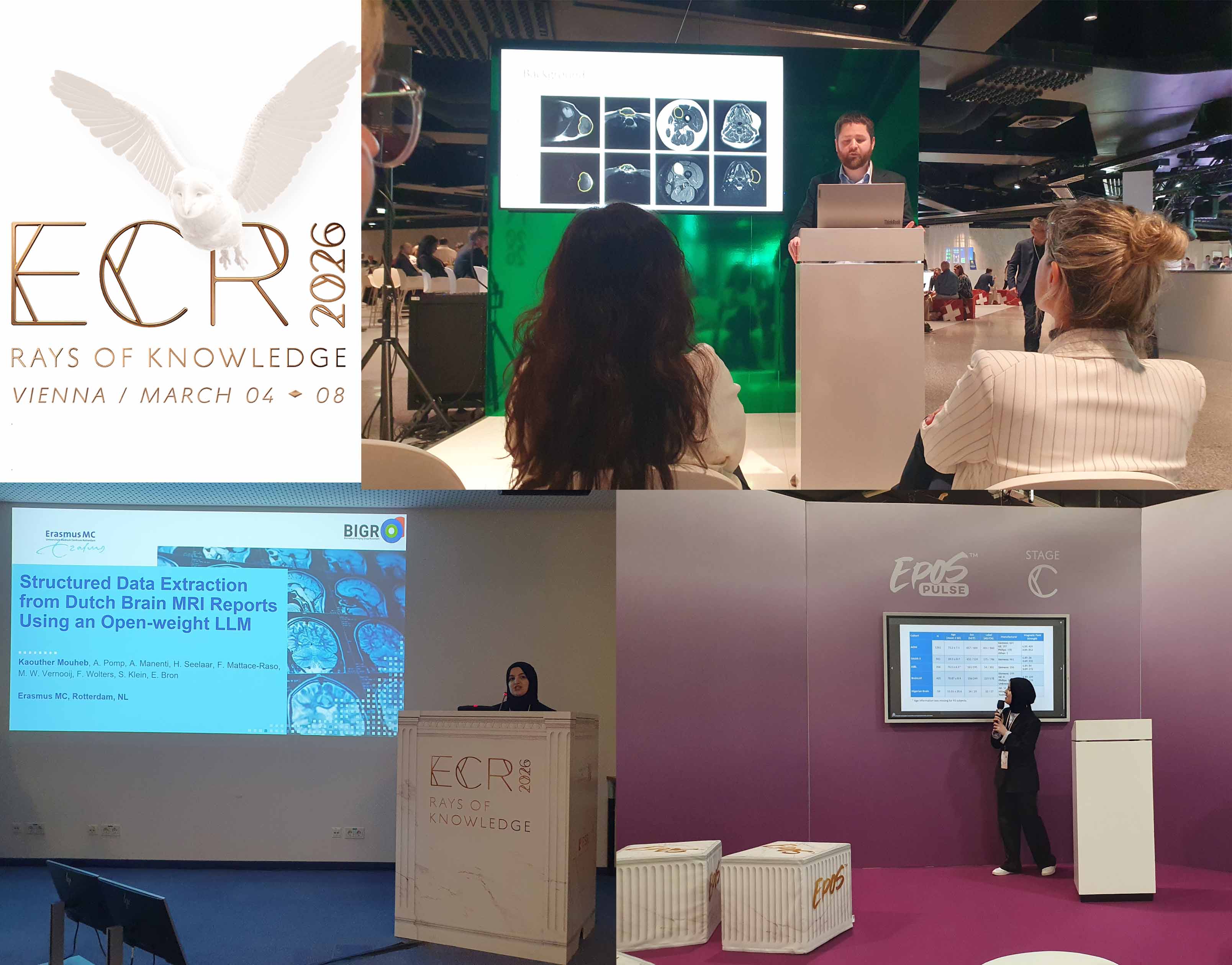
Several group members attended the European Conference on Radiology (ECR) in Vienna
February 15, 2026
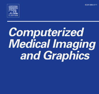
Wenjie Kang et al.’s paper, “An interpretable machine learning framework with data-informed imaging biomarkers for diagnosis and prediction of Alzheimer’s disease,” has now been officially published in Computerized Medical Imaging and Graphics!

February 1, 2026
The MICCAI 2026 Open Data Initiative, led by Martijn P.A. Starmans and Kaouther Mouheb from BIGR, has received a MICCAI Society Award for the Advancement of Health Equity.
Tumor localization and visualization using magnetic seed tracking and augmented reality
2024 - 2028
Imaging infrastructure for the COllaboration for New TReatments of Acute Stroke Consortium
2023 - 2028
The Rotterdam Study is a large population-based cohort designed to investigate causes and consequences of age-related diseases. Since 2005, brain imaging has been performed in over 6,000 participants using a 1.5T MRI scanner. A hardware upgrade in 2020 resulted in changes in signal reception across brain regions, introducing variability in subjects scanned directly before and after the scanner ugrade. This complicates longitudinal analysis, which is essential to study long-term disorders such as dementia. The aim of this project is to evaluate and apply feature-based harmonization methods (e.g., ComBat) and to explore deep learning-based approaches to correct for scanner-related variability in imaging biomarkers. Once harmonized, the data can be used to address longitudinal research questions, such as the association between cortical thickness and dementia risk. There is flexibility to explore specific research questions in the context of dementia. We are looking for a motivated student with an interest in neuroimaging analysis and epidemiology.
The aim of this project is to translate an existing deep learning framework for qMRI from synthetic data to real clinical MRI data. The student will retrain and validate the model using data from healthy volunteers, with the goal of moving qMRI closer to clinical acceptance.
The Carnegie staging system provides a standardized framework for assessing both normal and abnormal morphological development during the embryonic period. The Carnegie stages define 23 distinct steps that chronologically outline embryonic development from conception to the start of the fetal stage, indicating that all major organ systems are formed. Examples of these stages are illustrated in Figure 1. We have developed a deep learning model that can accurately predict the Carnegie stage of an embryo, given a 3D ultrasound image. Currently, the model gives no insight into which features were used to determine the stage. The aim of this project is to explore explainable deep learning methods or design novel features that explicitly assess developmental features such as neck curvature, limb position and brain ventricles in order to bring more insight into the models decision making. For more information contact: n.herrmann@erasmusmc.nl or w.bastiaansen@erasmusmc.nl.
Biomedical Imaging Group Rotterdam is a part of Erasmus MC